
USEFUL INFORMATION
In 1895, famous German physicist Wilhelm Roentgen discovered previously unknown rays. These rays penetrated various materials and soft tissues of the human body. Roentgen called them X-rays (a term still used in many countries). Later, in 1896, French scientist Henri Becquerel studying the properties of uranium salts discovered that they emit invisible rays. Following Becquerel, Marie and Pierre Curie discovered Polonium and Radium emitting similar rays. Thus, a new field of physics emerged, dealing with the study of ionizing radiation.
DATA TO BE INCLUDED IN RADIOLOGICAL STUDIES
In a broad sense, radiation is the transmission of energy in the form of waves or particles through a material medium. To clarify what is the form of radiation, how it affects the human body, etc., let us get acquainted with some concepts.
Atomic Structure, Isotopes
All matter consists of atoms. One can say that the entire mass of an atom consists of positively charged protons and neutral neutrons concentrated in its nucleus. Negatively charged particles called electrons move around the nucleus. Typically, atoms have an equal number of protons and electrons. The magnitude of the electric charges of protons and electrons is the same, but their signs are opposite. Therefore, atoms are electrically neutral.
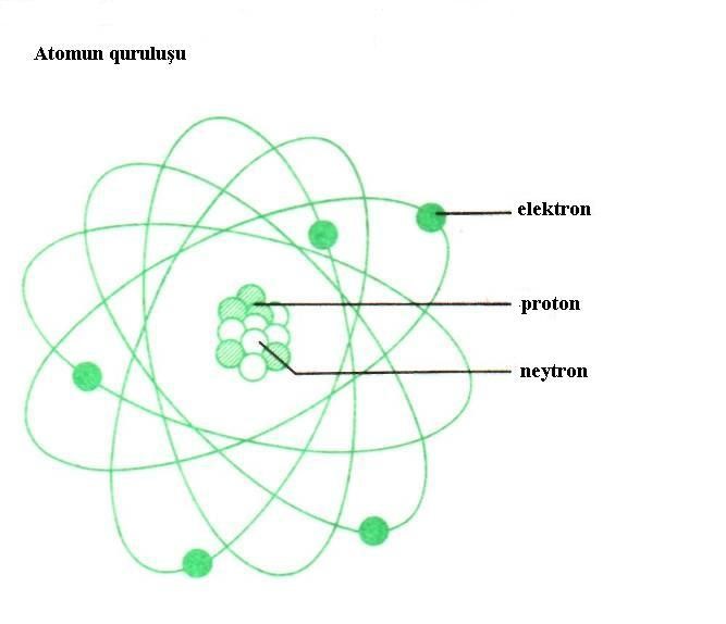
Some nuclides are stable and do not undergo any transformations in the absence of external impact. The vast majority of nuclides are unstable and decay spontaneously. Different nuclei release energy in different ways – in the form of electromagnetic radiation and/or particle flow. The spontaneous decay of unstable nuclides is called radioactivity. An unstable nuclide that decays by emitting radiation is called a radionuclide. To define the quantitative parameters of the radioactivity of any amount of radionuclide, the concept of activity is used. What is activity? Activity is the number of decays of any quantity of radionuclide per unit time and is measured in Becquerels (Bq). 1 Bq is the activity of a substance corresponding to 1 decay of that substance per second. The time for which the radionuclide activity is halved as a result of decay is called the half-life of that radionuclide. The type of emitted radiation, its energy, and the half-life of the nucleus are specific to each radionuclide. E.g., the half-life of the Uranium-238 and Bismuth-214 isotopes is, respectively, 4.47 billion years and 19.7 minutes. Ionizing radiation is radiation leading to the formation of ions and charged particles of different signs when interacting with the environment.
Ionizing Radiation Types
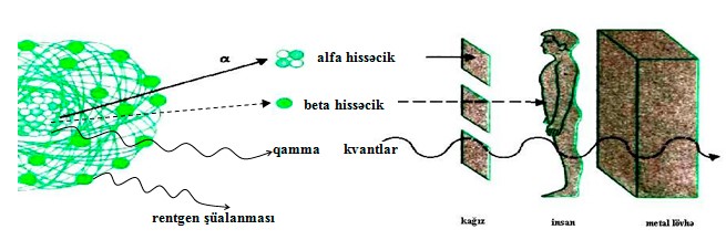
Alpha radiation consists of positively charged particles emitted by heavy nuclei of elements such as Uranium, Radium, Radon, and Plutonium. These particles are called alpha particles and consist of 2 protons and 2 neutrons. In fact, an alpha particle is a helium atom that has lost two electrons (doubly ionized), or the helium atom nucleus.
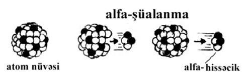
Alpha particles can travel only a few centimeters in the air and are completely absorbed by a regular sheet of paper. Alpha rays do not penetrate the skin. If any substance emitting alpha particles penetrates the body, it transmits energy to surrounding cells. Penetrating through the respiratory tract and open wounds, alpha emitters may cause harm to humans.
Beta radiation consists of electrons or positrons, which are much smaller than alpha particles. Electrons and positrons have negative and positive electrical charges, respectively.

Beta radiation can penetrate deeper and be absorbed by a metal plate or glass in its path, as well as ordinary clothing. Beta radiation can penetrate the germ layer of human skin, where new skin cells are formed. If beta emitters remain on the skin for a long time, they may cause skin damage.
Gamma radiation is electromagnetic waves. Gamma radiation particles are called gamma rays. Gamma rays have no electrical charge.
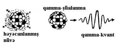
Gamma rays are capable of traveling long distances in the air, and their penetrating power is significant. High-density materials such as concrete or lead can be good barriers to gamma radiation.
There are two types of X-ray radiation: continuous and characteristic.
Continuous X-ray radiation arises as a result of the deceleration of electrically charged particles. E.g., when electrons attack a metal target in a vacuum (X-ray tube).
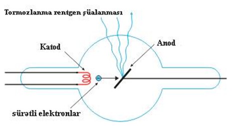
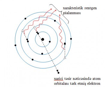
Characteristic X-ray radiation. An electron from the upper level (with higher energy) takes the place of another electron that has left its atomic orbital due to some impact. According to the same mechanism, the newly formed vacancy is occupied by an electron from a higher level. During each electronic transition, a photon is emitted with an energy equal to the energy difference between the corresponding levels. Since the energy levels (like the energies of the emitted photons) are specific to each atom and characterize it, the flux of emitted photons is called characteristic X-ray radiation.
X-rays can be blocked by high-density materials such as lead.
Neutron radiation consists of neutrons and is not ionizing as such. When a neutron hits a nucleus, it can activate the latter and release gamma rays or charged particles, thereby indirectly generating ionizing radiation. Another way for neutrons to indirectly cause ionizing radiation is by knocking out protons in hydrogen compounds due to collisions, which in turn generates proton ionizing radiation. Neutrons have a high penetrating power and can only be blocked by materials with low atomic weight such as water, paraffin, etc.
Radiation Dose
The harm caused by radiation to a living organism depends on the amount of energy transmitted to the tissue and the characteristics of the distribution of this energy in the tissue. Absorbed dose is the radiation energy absorbed by a unit mass of any substance. Its unit of measure is Gray. 1 Gray (Gy) is the dose at which 1 kg of a substance absorbs 1 J of energy.
Different types of radiation have different effects on the human body and tissues. The radiobiological effects caused by the same absorbed dose vary depending on the ionizing radiation type. This is due to different mechanisms of energy transmission when particles of various ionizing radiation types interact with tissue. To reflect the difference in the radiobiological effects of different radiation types, an equivalent dose concept has been introduced.
An equivalent dose is the product of the radiation dose absorbed in an organ or tissue by the corresponding quality factor (Table 1) of this radiation type. For photons (X-rays and gamma rays) and beta rays, this factor is 1, and for alpha rays, it is 20. This means that alpha radiation has 20 times the biological effect of beta radiation for the same amount of energy transmitted. These coefficients compare the biological effect of different radiation types with photon radiation or show the dose of absorbed photon radiation (X-ray, gamma radiation) in Grays, equivalent to that of 1 Gray of a given absorbed radiation (alpha, beta, neutron radiation) in terms of its biological effect.
Table 1. Average Values of Quality Factors
Type of radiation and energy range | k |
Photons of any energy | 1 |
Electrons and muons of any energy | 1 |
Neutrons with energy less than 10 keV | 5 |
10 to 100 keV | 10 |
100 keV to 2 MeV | 20 |
2 to 20 MeV | 10 |
20 MeV and more | 5 |
Protons with energy 2 MeV and more (except recoil protons) | 5 |
Alpha particles, fission fragments, heavy nuclei | 20 |
The unit of equivalent dose is Sievert (Sv).
Organ or tissue | wT | Organ or tissue | wT |
Reproductive glands | 0.20 | Liver | 0.05 |
Stomach | 0.12 | Esophagus | 0.05 |
Bone marrow (red) | 0.12 | Thyroid | 0.05 |
Lungs | 0.12 | Bone surface cells | 0.01 |
Colon | 0.12 | Skin | 0.01 |
Mammary gland | 0.05 | Other | 0.05 |
Bladder | 0.05 | Total: | 1 |
Table 2. Weighting Factors for Organs and Tissues
Organs and tissues of the human body have different radiosensitivity. Thus, the same dose of the same radiation type, absorbed in different organs and tissues, may cause different biological effects depending on the characteristics of those tissues. To consider this, the effective dose concept is introduced, which is determined by multiplying (the product of) the weighting factor of the corresponding organ or tissue (Table 2) by the equivalent dose absorbed by that organ or tissue and summing the values obtained for all organs and tissues. Weighting factors are defined empirically, provided that their sum for the entire body does not exceed 1.
Since weighting factors do not have a unit, the effective equivalent dose is measured in Sieverts as the equivalent dose.
In contrast to the equivalent dose, the effective equivalent dose characterizes the likelihood of a radiological effect for the entire body and not individual organs or tissue.
Background Radiation, Exposure Dose
Background radiation is the dose rate of ionizing radiation from natural sources of cosmic and terrestrial origin, as well as artificial radionuclides scattered in the biosphere as a result of human activity.
Background radiation remains relatively constant and affects all life on Earth.
Background radiation has the following components:
- a) natural background radiation – ionizing radiation from natural sources of cosmic and terrestrial origin;
- b) technologically enhanced natural background radiation – ionizing radiation from natural sources certainly changed as a result of human economic activity;
- c) artificial background radiation is caused by nuclear weapons testing, nuclear energy waste, and radiation accidents.
The exposure dose is measured in Coulombs per kilogram (C/kg) or Roentgens (R) based on the air ionization caused by radiation. 1 Roentgen is the dose at which X-ray or gamma radiation generates 2.08×10⁹ ion pairs in the air under normal conditions.
The unit of exposure dose rate is Roentgen/hour.
In the Republic of Azerbaijan, the natural background radiation is up to 20 µR/h.
According to regulatory documents on radiation safety, the following dose limits are set for the population’s exposure above the natural background radiation.
Dose Limits for the Public under Normal Conditions
- a) annual effective dose – 1 mSv/g;
- b) annual effective dose to the eye lens – 15 mSv/g; and
- c) annual effective dose to the skin – 50 mSv/g.
Radiation Dose Limits for the Public under Specific Conditions
Under specific conditions, annual doses received by appropriate critical groups of the population should not exceed 5 mSv for any single year, provided that the average annual dose received over 5 consecutive years does not exceed 1 mSv.
Documents Regulating Radiation Safety of the Population
Law No. 423-IQ of the Republic of Azerbaijan On Radiation Safety of the Population, dated December 30, 1997
Law No. 371 of the Republic of Azerbaijan On Sanitary and Epidemiological Well-being, dated November 10, 1992
Radiation Hazard Symbol
If you find any unknown item bearing a radiation hazard symbol, please immediately report it to 112 Service of the Ministry of Emergency Situations.
